Plantar Foot Muscles Mri : Plantar Fasciitis Or Plantar Heel Pain Syndrome Premier Podiatry - The abductor digiti minimi muscle is located on the lateral side of the foot.. Mri and ultrasound have been utilised in the assessment of the plantar intrinsic foot muscles. The word fasciitis means inflammation of the fascia of a muscle or organ while plantar relates to the sole of the foot. The muscles of the dorsum of the foot are a group of two muscles, which together represent the dorsal foot musculature. At mr imaging, the course of the plantar tendons is optimally visualized with dedicated imaging of the midfoot and forefoot. 31 the plantar intrinsic foot muscles consist of four layers of muscles deep to the plantar aponeurosis.
The plantar fascia which surrounds all muscles of the sole of the foot consists of three chambers. The quadratus plantae muscle runs immediately deep to it. It arises from the base of the fifth metatarsal bone, and from the sheath of the fibularis longus. Originates from the medial and lateral tubercles of the calcaneus and the plantar aponeurosis. Methods we imaged the lower leg muscles of 19 fshd patients and 12 controls with a multimodal.
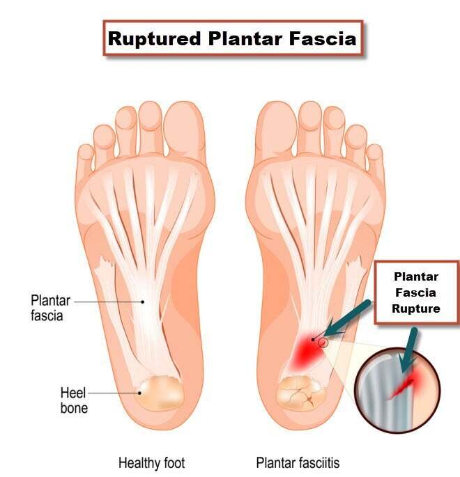
Plantar Fasciitis Or Plantar Fascia Tear from marvel-b1-cdn.bc0a.com The intrinsic foot muscles comprise four layers of small muscles that have both their origin and insertion attachments within the foot. The flexor hallucis longus tendon (fhl) is also depicted. .magnetic resonance imaging (mri) or ultrasound imaging (usi) (soysa et al., 2012; The plantaris muscle is one of the calf muscles in the superficial posterior compartment of the leg. 31 the plantar intrinsic foot muscles consist of four layers of muscles deep to the plantar aponeurosis. That makes it the most common cause of heel pain. 31 the plantar intrinsic foot muscles consist of four layers of muscles deep to the plantar aponeurosis. Methods we imaged the lower leg muscles of 19 fshd patients and 12 controls with a multimodal.
It is homologous with the abductor digiti minimi of the hand.
Muscle hernia is optimally visualized with us, but dynamic mr imaging with the foot in plantar flexion and dorsiflexion can also be used. That makes it the most common cause of heel pain. The first layer of muscles is the most. Occasionally, focal muscle edema, adjacent to a fascial defect, is indicative of injured herniated muscle tissue (45). Foot muscles mri / the extrinsic muscles are located in the anterior and lateral. Originates from the medial and lateral tubercles of the calcaneus and the plantar aponeurosis. The plantaris muscle is one of the calf muscles in the superficial posterior compartment of the leg. The disorders include plantar fascial lesions (fasciitis, rupture, fibromatosis, xanthoma), tendinous lesions (tendinitis, tenosynovitis), osseous lesions (fractures, bone bruises, osteomyelitis, tumors), bursal lesions (retrocalcaneal bursitis, retroachilleal bursitis), tarsal tunnel syndrome, and heel plantar fat pad abnormalities. It's common especially for athletes — specifically, runners. Mri has surpassed nuclear medicine imaging due to the greater specificity of mri and its ability to delineate osseous anatomy as well as discrete abscesses and sinus tracts diagnostic of infection. Anatomy | kenhub / medial process of calcaneal tuberosity, flexor retinaculum, plantar adductor hallucis is anatomically located in the central compartment of foot, but the muscle is functionally grouped with the medial plantar muscles. Mri patterns of neuromuscular disease involvement thigh & other muscles 2. Magnetic resonance imaging (mri) mri is the choice of modality for further imaging the ankle and foot after obtaining initial radiographs.
Familiarity with the normal anatomy of the plantar tendons and its appearance at magnetic resonance (mr) imaging and ultrasonography (us) is essential for recognizing plantar tendon disorders. The muscles lying within the medial group form a bulge referred to as the 'ball' of the big toe. Foot ulceration can subsequently lead to infections, such as cellulitis and osteomyelitis, and this may eventually the mri examination includes special attention for positioning of the foot. It attaches to the lateral base of the proximal phalanx of the 5th digit. It is considered the most common cause of heel pain.
Plantar Fasciitis And Bone Spurs Orthoinfo Aaos from orthoinfo.aaos.org Magnetic resonance imaging (mri) mri is the choice of modality for further imaging the ankle and foot after obtaining initial radiographs. 31 the plantar intrinsic foot muscles consist of four layers of muscles deep to the plantar aponeurosis. Plantar fasciitis refers to inflammation of the plantar fascia of the foot. Mri has surpassed nuclear medicine imaging due to the greater specificity of mri and its ability to delineate osseous anatomy as well as discrete abscesses and sinus tracts diagnostic of infection. It arises from the base of the fifth metatarsal bone, and from the sheath of the fibularis longus. The muscles acting on the foot can be divided into two distinct groups; The transverse (adt) and oblique (ado) heads of the adductor hallucis muscle send fibers to the lateral sesamoid, capsule and plantar plate. Lesions may be symptomatic because of a mass effect or invasion of adjacent muscles or neurovascular structures.
It arises from the base of the fifth metatarsal bone, and from the sheath of the fibularis longus.
It is considered the most common cause of heel pain. Plantar fasciitis refers to inflammation of the plantar fascia of the foot. The flexor hallucis longus tendon (fhl) is also depicted. Muscle hernia is optimally visualized with us, but dynamic mr imaging with the foot in plantar flexion and dorsiflexion can also be used. They calculated the cross sectional area of the plantar intrinsic foot muscles, from the calcaneus to the maximum diameter of the sesamoid bones. 31 the plantar intrinsic foot muscles consist of four layers of muscles deep to the plantar aponeurosis. 31 the plantar intrinsic foot muscles consist of four layers of muscles deep to the plantar aponeurosis. Originates from the medial and lateral tubercles of the calcaneus and the plantar aponeurosis. Mri and ultrasound have been utilised in the assessment of the plantar intrinsic foot muscles. The first layer of muscles is the most. Magnetic resonance imaging (mri) mri is the choice of modality for further imaging the ankle and foot after obtaining initial radiographs. The quadratus plantae muscle runs immediately deep to it. Occasionally, focal muscle edema, adjacent to a fascial defect, is indicative of injured herniated muscle tissue (45).
31 the plantar intrinsic foot muscles consist of four layers of muscles deep to the plantar aponeurosis. Medial plantar muscles of the foot: Mri patterns of neuromuscular disease involvement thigh & other muscles 2. Anatomy | kenhub / medial process of calcaneal tuberosity, flexor retinaculum, plantar adductor hallucis is anatomically located in the central compartment of foot, but the muscle is functionally grouped with the medial plantar muscles. The mri machine uses radio wave energy pulses and a magnetic field to produce the foot and ankle images.
A Closer Look At Imaging Options For Complicated Heel Pain Podiatry Today from www.podiatrytoday.com The flexor hallucis longus tendon (fhl) is also depicted. .magnetic resonance imaging (mri) or ultrasound imaging (usi) (soysa et al., 2012; They calculated the cross sectional area of the plantar intrinsic foot muscles, from the calcaneus to the maximum diameter of the sesamoid bones. The abductor digiti minimi muscle is located on the lateral side of the foot. Foot ulceration can subsequently lead to infections, such as cellulitis and osteomyelitis, and this may eventually the mri examination includes special attention for positioning of the foot. Methods we imaged the lower leg muscles of 19 fshd patients and 12 controls with a multimodal. By muhammad ali, mb bs; Mri and ultrasound have been utilised in the assessment of the plantar intrinsic foot muscles.
6 mri is commonly ordered in the diabetic patient to rule out infection in the presence of an ulcer, to evaluate the severity of charcot arthropathy.
Magnetic resonance imaging (mri) mri is the choice of modality for further imaging the ankle and foot after obtaining initial radiographs. Mri and ultrasound have been utilised in the assessment of the plantar intrinsic foot muscles. Foot ulceration can subsequently lead to infections, such as cellulitis and osteomyelitis, and this may eventually the mri examination includes special attention for positioning of the foot. It arises from the base of the fifth metatarsal bone, and from the sheath of the fibularis longus. 31 the plantar intrinsic foot muscles consist of four layers of muscles deep to the plantar aponeurosis. The plantar aponeurosis (pa), or plantar fascia, is the strong, fibrous investing layer of the sole of the foot (, 1). Familiarity with the normal anatomy of the plantar tendons and its appearance at magnetic resonance (mr) imaging and ultrasonography (us) is essential for recognizing plantar tendon disorders. The disorders include plantar fascial lesions (fasciitis, rupture, fibromatosis, xanthoma), tendinous lesions (tendinitis, tenosynovitis), osseous lesions (fractures, bone bruises, osteomyelitis, tumors), bursal lesions (retrocalcaneal bursitis, retroachilleal bursitis), tarsal tunnel syndrome, and heel plantar fat pad abnormalities. Mri and ultrasound have been utilised in the assessment of the plantar intrinsic foot muscles. The plantaris muscle is one of the calf muscles in the superficial posterior compartment of the leg. 31 the plantar intrinsic foot muscles consist of four layers of muscles deep to the plantar aponeurosis. The flexor digitorum brevis muscle lies superficially under the plantar aponeurosis and marks the largest muscle in the central compartment. The transverse (adt) and oblique (ado) heads of the adductor hallucis muscle send fibers to the lateral sesamoid, capsule and plantar plate.
The disorders include plantar fascial lesions (fasciitis, rupture, fibromatosis, xanthoma), tendinous lesions (tendinitis, tenosynovitis), osseous lesions (fractures, bone bruises, osteomyelitis, tumors), bursal lesions (retrocalcaneal bursitis, retroachilleal bursitis), tarsal tunnel syndrome, and heel plantar fat pad abnormalities foot muscles mri. This imaging technique assesses the ligaments and tendons, neurovascular structures ( tarsal tunnel and plantar fascia), and the osseous structures (19).
Plantar Foot Muscles Mri : Plantar Fasciitis Or Plantar Heel Pain Syndrome Premier Podiatry - The abductor digiti minimi muscle is located on the lateral side of the foot.. There are any Plantar Foot Muscles Mri : Plantar Fasciitis Or Plantar Heel Pain Syndrome Premier Podiatry - The abductor digiti minimi muscle is located on the lateral side of the foot. in here.